Precise, Repeatable Imaging Behind the Iris
The ArcScan Insight® 100 instrument provides anatomical detail to make informed, clinically sound treatment and surgical decisions. With the images you capture, you can visualize and measure in microns areas of the eye that are simply not clearly visible with other devices.
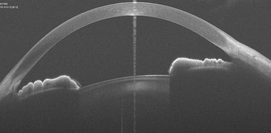
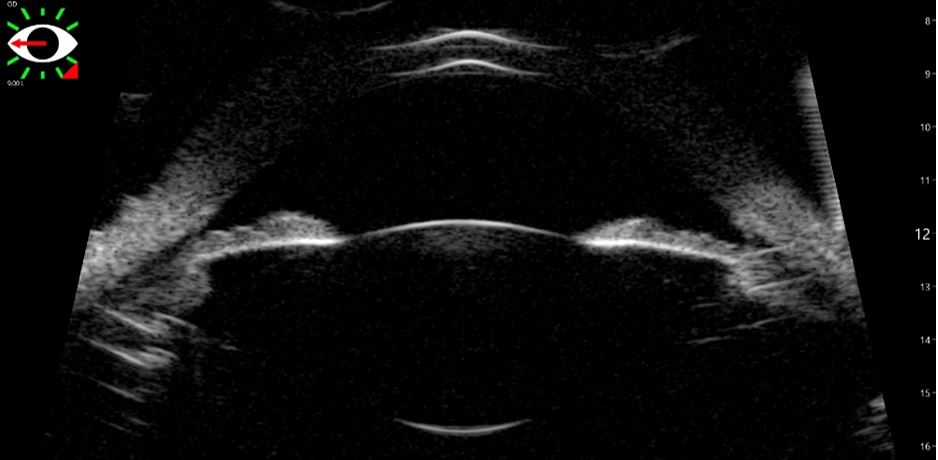
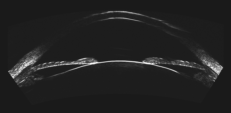
The Insight 100 moves in an arc, perpedicular to the curvature of the anterior ocular surface. Using ultra-high-frequency ultrasound, it penetrates the eye’s anatomy, including anterior chamber depth, angle-to-angle width, and sulcus-to-sulcus width, and pathologic structures, such as solid masses and cysts.
The result? Images that elicit comments like “I will simply not do ICL surgery without it.”

The ArcScan Insight® 100 uses proprietary technology and flexible software to allow you to efficiently capture scan sets as well as tailor the meridians and depth of the Z axis. With years of software development, the scan sets offer options like epithelial mapping, Kerataconus risk identification, ICL sizing data, and coronal scans. In addition to a hands-on training, our Clinical Applications team and peer resources help you best utilize the technology for your clinic and patients.
Patient-Centric Design
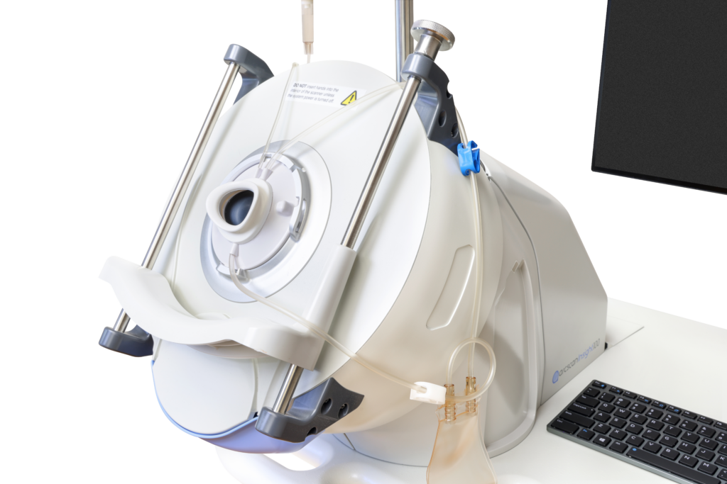
The device is designed to comfortably position the patient so their eye looks through the center of the disposable EyeSeal while keeping their head positioned well. EyeSeal disposables are used to prevent cross-contamination between patients.
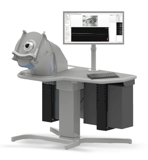
A dramatically new approach to ultrasound imaging, the device was created with efficiency and ease of use in mind. It comprises four main components: the ArcScan instrument module, the Insight software package, the fluidics module, and the electronics unit.
Clinical Benefits
In contrast to handheld ultrasound devices, the patient is comfortably seated in front of the device, easily accessible to the operator next to them. By adjusting the table and guiding the patient as they lean in, the operator uses visual feedback on screen to center the eye. Then, in mere seconds, each scan can be quickly completed.
Seat time may vary, but experienced operators report 5 to 10 minutes is typical.
Since each practice is unique, a conversation with ArcScan staff is a great next step.
UBM vs Insight®100
OCT vs Insight®100