Corneal tomography and imaging uses:
- Early Detection of Keratoconus Risk
- Pre- and Post-LASIK
- Trans Epithelial PRK Biometry
- Evaluation of Corneal Scars
In addition to the Corneal Scan Set, the operator can zero in on areas of concern, adjusting the depth and meridians to pinpoint anatomy to explore. With the ArcScan, early detection of keratoconus risk (or not) means the clinician can confidently move ahead with full anatomical knowledge to select the most appropriate type of care.
We can achieve a very precise image of your eye that provides an accurate assessment of the type of vision correction that will be the best prescription for your personal eye care.
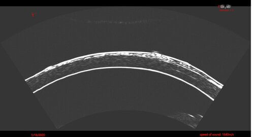
Using Ultrasound to Create Corneal Imaging
The ability to image the entire anterior segment in minute detail shows the actual anatomical structure.
Studies have shown that having this data available may enable clinicians to increase their LASIK and Trans Epithelial PRK candidacy and rule in patients from LASIK and Epithelial PRK surgery due to suspicious front and back surface corneal tomography 1,2
Professor Dan Reinstein, MD
How the Insight 100 Has Changed His Practice
How it Works
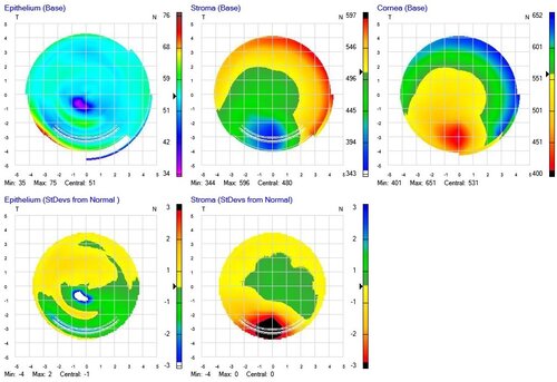
The Corneal Imaging Mode uses an arc radius of 8-9 mm to match the corneal radius of curvature, capturing the entire cornea using four equally spaced meridians. The Insight 100’s ability to take precise corneal tomography and micro-measurements in the cornea and anterior segment provides early detection of subtle keratoconus eye disease, allowing the patient to receive the best care.
ArcScan’s tomographic micro-measurements ocreate an epithelial thickness map of the entire 10mm diameter of the cornea. This produces a deeper understanding of the corneal layers that evaluates potential Keratoconus and other aberrations.