
“Since we’ve gotten the ArcScan I can with confidence implant the ICL and know that the vault is going to be perfect every time.”
Dr. Tom Tooma, Chief Medical Officer
NVISION Eye Center
Middle Segment Ultrasound Imaging
The ability to precisely determine pIOL lens sizing and placement impacts patient outcomes and clinic efficiency – and much more. The data from the Insight 100’s integrated measurement tools and Anterior Segment scans is designed to support your decisions for pIOL lens sizing and placement. Then, check your outcomes with a post-operative scan.


Just Right
As you plan for and review pIOL surgery, zeroing in on the sizing and anatomy behind the iris leads to outcomes success. The Insight 100 pIOL Scan Sets capture anatomy that is easily measured using the Rapid Caliper Tool. With the data you gather, nomogram results are demonstrably more precise than the traditional sulcus-to-sulcus measurement.1 With further interpretation, you can identify pathologies or anatomical challenges to make surgical planning more comprehensive.
By employing the Insight 100 and its behind-the-iris ultrasound imaging, you and your patients can count on the precision and repeatability to prepare for and measure the outcomes of pIOL surgery.1
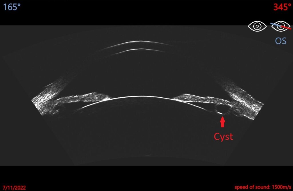
Anatomy and Artifacts
The Insight 100 Anterior Scan is comprised of tailored meridians, with very-high-frequency ultrasound multiple sweeps to deliver images that helps Ophthalmologists focus on elements like:
- Scleral spur
- Anterior chamber
- Ciliary body
- Masses and cysts
- Irido-corneal angle
- And more
Interested in learning more? Contact us to request a call or get more information.